The idea that viruses "don't exist" is a very flamboyant statement. It is bound to turn people off that you're just one of those crazy, dumb, "conspiracy theorists". However, let's investigate, scientifically, further into this statement. First of all, one thing to clarify, is that diseases that are typically purported to be CAUSED by viruses, certainly exist. No one denies that people get sick and die from COVID, measles, smallpox, etc. These disease processes exist. The contention is that they are caused by a particle known as a virus, that spreads from organism to organism via the mechanisms of causing cells to lyse (burst), releasing copies of the virus. While I am going to go into my presentation of the topic, this is a very thorough
Before we get into analyzing the scientific studies that purport to prove that viruses exist and are causal in disease, we should look at some interesting studies undertaken in the early 1900s. This describes a study that took place among 68 army men with no history of infection from the spanish flu. Keep in mind, this is the demographic that was the most whiped out by the spanish flu source. Here is another such study using 16 men. They took nasal secretions and sputum from people sick with the spanish flu, and inoculated them into the test subjects, and none got sick. They then had the test subjects converse with people that were sick and in fact get coughed on in the face by them, and none got sick. They then took sputum and blood from sick people and injected them subcutaneously into the subjects. None got sick.
Max Joseph von Pettenkofer was a popular father of the sanitation movement in Germany in the late 1800s. He was a rival of Robert Koch, the father of germ theory and namesake of the Robert Koch Institute, and he did not believe that bacteria were causal in disease. He believed this so much that he in fact obtained a sample of bouillon from Robert Koch that had a large dose of cholera and "consumed the bouillon in a self-test in the presence of several witnesses on 7 October 1892. He also took bicarbonate of soda to neutralise his stomach acid to counter a suggestion by Koch that the acid could kill the bacteria. Pettenkofer suffered mild symptoms for nearly a week but claimed these were not associated with cholera."
Dr. Robert Willner, at 40:00 in this video, inoculates himself with the blood of an HIV positve man to set out to prove that HIV does not cause AIDS. Becoming extremely famous in Spain. He speaks about it in an excerpt from his book The Beginning of the End. The eery thing about this case is that, I had read online that he died 6 months later from a heart attack. You can see this on his wikipedia page. However, there is only 1 source given, and it links to an extremely old website with no source for the claim. It is claimed elsewhere that he died of a car crash. You can check yourself by googling and trying to find an obiturary or some proof of his death, or anything past 1994, but you will find nothing. He probably did not die from AIDS, because if he had this would be news plastered everywhere for the HIV=AIDS movement. Plus, not even sources that are anti-willner say it was aids, but a "heart attack". Also, even by mainstream dogma, HIV should not kill in the span of 3-6 months. One thing seems clear. We don't know how he died, and may never find out. The conspiracy minded people will say that he was "taken out". I'll let you decide.It's important to address these one by one. In influenza studies, while they are very surprising to many, they of course don't necessarily mean that the flu is not contagious. However, germ theorists had to alter their theory in the face of these findings. This is where the concept up the immune system began to take place to explain why people can not get sick from the agents that a priori are assumed to be disease causing. The problem I have with this reasoning, however, is that it is really unfalsiable in face of observational evidence. If someone is exposed to a virus and they don't get sick, it's because they are immune. If they are exposed and they sick, it's because it is contagious. It is a non falsiable premise. However, with all this said, it would seem statistically unlikely for this to be the case given that these people had 1) not previously been exposed and 2) were in the exact demographic that were dying in droves from this virus, exposed not only through natural means but through completely unnatural means (subcutaenous inoculation).
In the case of the doctor drinking cholera juice, we see that he did not become sick. It is stated that he experienced short lived "minor symptoms", which if we are to assume that this is true could easily just be explained by his consumption of the sodium bicarbonate (depending on the quantity), some unknown entity in the bouillon he drank, or just possibly a placebo affect. I've also read reports that he did not experience any sypmtoms, so the severity of symptoms seems to range from non-existent to minor. In any case, this would seem extremely unlikely given a germ theory being correct and the bacteria itself causing disease. But it is not a rigorous scientific experiment rife with flaws, just interesting to point out.
Finally, we have the inoculation of blood from Dr. Robert Willner. Unfortunately as was mentioned, we were not able to see the long term results of this, but I thought it was another interesting instance of self testing to bring up. While these are not my groundbreaking examples of why viruses do not exist, I think they are useful to keep in mind, and to also keep in mind that there are no human studies that have ever been able to prove contagion. In modern day this study would never be undertaken due to medical ethics, but it is interesting to see that when we had much more lax rules of medical ethics, some of these experiments were undertaken on humans and they came back negative.
We can also see animal experiments trying to prove transmission of viruses. The most egregious, and barbaric of which are the experiments done on monkeys to prove the transmissions of polio. These attempts to prove the transmission of polio involved drilling into monkeys skulls and injecting large quantities of spinal matter from a child diagnosed with polio. While they tried to infect them via normal routes, i.e. through the mouth, and even injecting subcutaenously in the arm, they were unable to show any paralysis or damage. However, when they injected directly into the brain their was neurological damage. A truly groundbreaking discovery. The problems with this should not need to be stated but are numerous. Number 1, they make no attempt to isolate any disease causing agent in the solution of spinal cord mixture from the child, so one could not be sure that a virus is the cause of the issue or some other cellular debris or toxic. Number 2, there was no control monkey that was injected, say saline or water, into the skull to see if there were similar findings, because it seems to me that one would EXPECT serious reactions to having your skull split open and foreign liquids injected into it. But this was the state of groundbreaking virological science in the early 1900s.
You can read more about the attempts to prove the transmissibility of polio, and you will find the same results, that they had an extremely difficult time proving that it was contagious at all. This will be a recurring theme in virology. When presented with evidence that their theory may be wrong, instead of considering that fact, they assume that they simply need to try harder and double down. They were unsuccessfully able to make rabbits, guinea pigs, mice, or many other animals sick through inoculations and normal routes of exposure, and their conclusion was that we just need expose them more invasively. So they inject extremely large amounts into the brain or stomach cavity, with no control experiments. Incredible.
But again, all these are not necessarily proving that viruses do not exist. After all, we have not actually tried to isolate, image, and study one of these particles. It's worth keeping in mind that when the viral hypothesis was invented in the late 1800's, we did not have the technology to actually image them. They are in the nanometer range, and therefore are not able to be seen via traditional light microscopes. It wouldn't be until the 1930's that the Electron Microscope was invented and we could attempt to image things on that small scale. So surely, since that point, a virus has been isolated and imaged, no?
Let's take the most recent example of SARS-COV-2. We can take a look at this study, that purports to have "isolated" SARS-COV-2. We can look at figure 1 image D, and see from the description "Electron microscopic image of the viral isolate". However, it is obviously not an isolate as we can see these particles budding from a cell with plenty of other structures. They claim to have "isolated" the virus via nucleic acid extraction of the (unpurified) sample, but since the SARS-COV-2 genome has been sequenced from a similarly unpurified and unisolated sample, it is slightly circular reasoning.
I know that may have been a lot, so let me explain further what is done in this study (in plain English), along with all other viral isolation studies since John Enders in 1952. A sample from a sick person is taken, for example the sputum (lung fluid) in the case of COVID. This sample will either go through no filtering steps, or it will be centrifuged, which will basically accomplish getting out any particles that are larger than the nanometer range. This gets rid of bacteria and leaves you with things the size of viruses, proteins, and other macromolecules. So what we have at this point is an unisolated mixture from a sick person that may have any array of nucleic acids, proteins, viruses, molecules, toxins, etc. of unknown origin. They will then take a culture of Vero cells (monkey kidney cells) in a petri dish (in vitro), and add a mixture of antibiotics, a nutritive medium (barely enough to sustain life), and calf serum (components of calf blood). They then place the unpurified sample from the sick person into this petri dish, and they wait to observe Cyptopathic Effects (CPE), or cellular damage. Once the CPE is noticed, they conclude that the virus must be the cause of this damage.
The final step they take in this paper, although they rarely actually do this in most virology papers, is to take an electron microscope image of the "virus". The problem is that there has been no attempt to isolate a viral particle from any other particle, and this is what I alluded to earlier. You can see in the image that "COVID" is seen to be budding off of a larger body, which is the cell. So this is not an isolated solution, it is a solution in cell culture. Furthermore, in terms of proving that the virus is causal in disease, besides the fact that they did not isolate the virus first which would be necessary to prove that it and it alone caused the CPE, they do not use a control cell culture! And this has never been properly done in virology. A control cell culture would be to have a cell culture in which you use the monkey kidney cells, cultured with antibiotics, nutritive medium, and calf serum in the same amounts, and do NOT add the "viral" mixture. Then, if you were to observe CPE in the control culture, you would know that whatever you added to the mixture did not actually cause the damage. And this is what we would expect to see because antibiotics and starving of cells in vitro are known to cause cell death!
In fact, here is a paper that shows that antibiotics induce the release of a type of cellular debris known as "exosomes". Exosomes are extracellular vesicles, that consist of a protein that engulfs nucleic acids for transport. Sound familiar? If you compare images of exosomes to the "COVID" images above, you see they look indistinguishable. This is not just me "realizing" this, this is known in mainstream science. This mainstream virological paper says that "The remarkable resemblance between EVs and viruses has caused quite a few problems in the studies focused on the analysis of EVs released during viral infections. Nowadays, it is an almost impossible mission to separate EVs and viruses by means of canonical vesicle isolation methods, such as differential ultracentrifugation, because they are frequently co-pelleted due to their similar dimension [56,57]. To overcome this problem, different studies have proposed the separation of EVs from virus particles by exploiting their different migration velocity in a density gradient or using the presence of specific markers that distinguish viruses from EVs [56,58,59]. However, to date, a reliable method that can actually guarantee a complete separation does not exist." In other words, we have never actually been able to separate "viruses" from exosomes. In other words, what we are looking at in those covid images are exosomes.
We see another paper here showing that we were seeing covid like particles in patients before the pandemic even began. Here is another paper on the same issue published on the CDC's website. We actually see a similar problem in papers on measles virus "isolation" as well. Here we see another such paper. Here we see an admission that "Ruckle has lately reported similar findings and in addition has isolated an agent from monkey kidney tissue that so far is indistinguishable from human measles virus. The problem, however, of the origin of the agent responsible for the presence of these antibodies in apparently normal monkeys has not yet been solved. There is a potential risk in employing cultures of primate cells for the production of vaccines composed of attenuated virus, since the presence of other agents possibly latent in primate tissues cannot be definitely excluded by any known method." In plain english, the "measles virus" is found in the monkey cell cultures used in complete absense of any viral infection. You can see another paper attempting to isolate COVID using the same methodology here.
Another point to add is that, even if all of these flaws in these studies had been fixed, and a pure isolation of a virus was maintained, and a control culture used, there would still be problems with the conclusion that these particles cause viral illnesses in humans, as admitted by John Enders in the paper that landed him a nobel prize in virology. source "While there is no ground for concluding that the factors in vivo are the same as those which underlie the formation of giant cells and the nuclear disturbances in vitro, the appearance of these phenomena in cultured cells is consistent with the properties that a priori might be associated with the virus of measles." What he is saying here is that, you cannot necessarily extrapolate things that you see in vitro (in the petri dish), with what you may see in a whole organims (in vivo). In the same paper, Enders admits that "As control antigens, fluids were taken from uninoculated cultures maintained under the same conditions as well as fluids from cultures of the agent producing changes superficially similar to those caused by the measles agents and which are mentioned below". In other words, Enders saw the same cytopathic effects in the uninfected cell cultures as he did with the "measles" virus. He did a proper control!
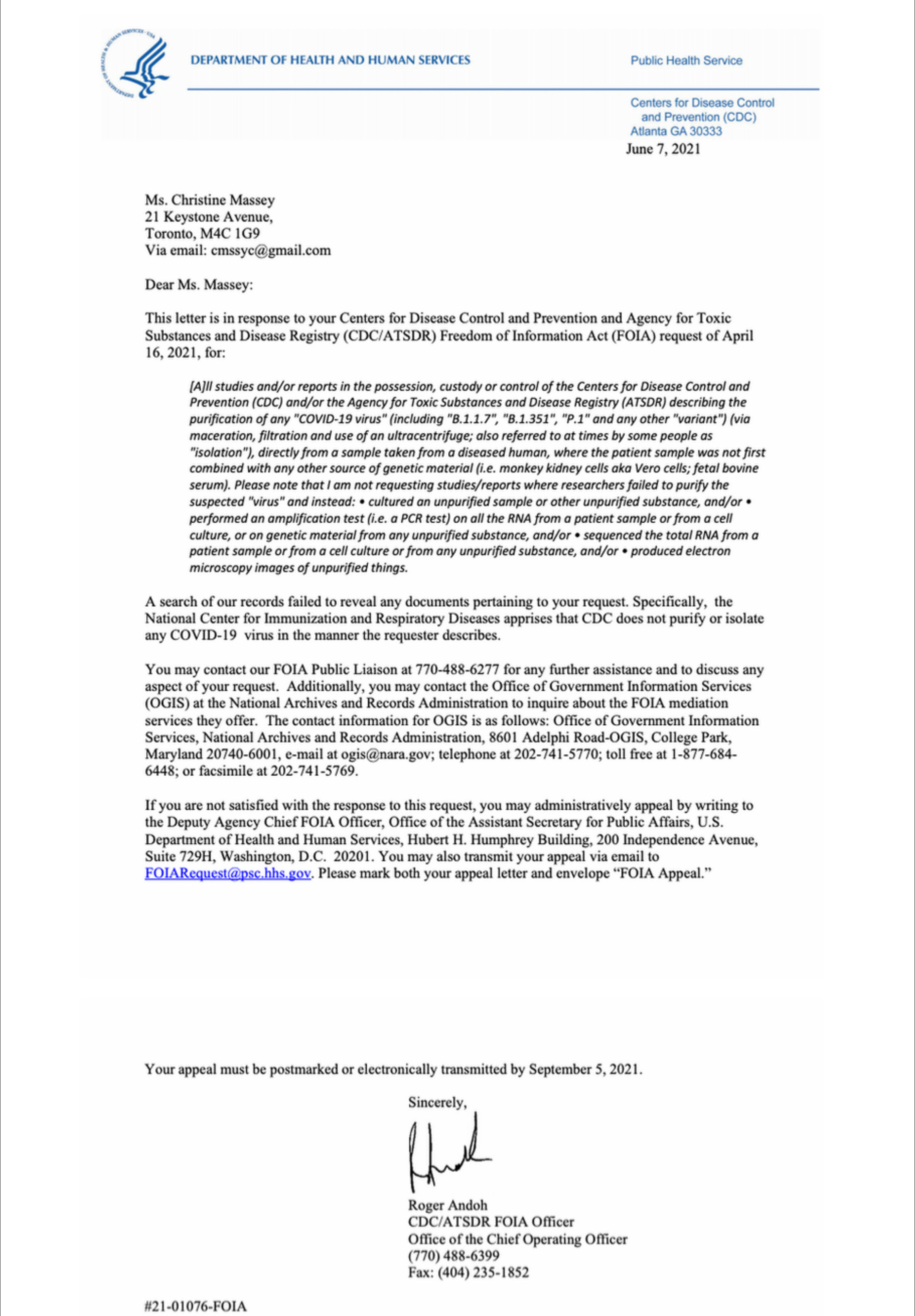
You may be thinking, ok this all makes sense but how can I trust your judgement? After all, most scientists agree that COVID exists, they can't all be wrong right, lying, or incompetent, right? I would encourage you to ask professionals and virologists yourself. In fact, that is what the author of this article did. He asked the authors of all the studies that purport to isolate the COVID virus the simple question "Do your electron micrographs show the purified virus?" They all responded no. There is also this FOIA (Freedome of Information Act) from the CDC that concedes that they have not isolated a COVID-19 particle.
CORMAN-DROSTEN REVIEW REPORTAntibody titers used to compare antibody levels pre-and post pandemic. Need to look into titer methods used
However, no attempts to prove causation in disease or that they are in fact viruses.
SARS-Cov-1No monkeys died, euthenized after 6 days, given ketamine before death. Damage to organs were determined, no control group. Viral pictures taken with no isolation.
This paper claims to use a "mock infected" control cell line with no CPE. However, no specific discussion or methods are stated about how the control cell lines were prepared and its mentioned in passing only once.
Based on this article, they state that "Confluent cells were infected with SARS-CoV, which resulted in a multiplicity of infection of 1.7 (results not shown) or were mock-infected with medium only." So while there is very little discussion on the mock cultures, they at least admit that they did no add any antibiotics to the control cell lines... This casts some doubt on the methods of the previous paper, and the only way to confirm would be to contact the authors most likely.
"Schmidt 3 from a series of 196 filtrate inoculations from 16 different subjects suffering with coryza concludes that his investigations do not support the filtrable virus theory of "colds." Branham and Hall 4 state that in attempting to cultivate filtrable viruses from nasopharyngeal secretions in colds and influenza on the various mediums employed by them, no bodies were found in the cultures which could not be found also in those from normal persons, in controls, in all simple mediums examined, and on blank slides. This work does not support the work of Foster," who contends that definite bodies were found when cultivated according to Noguchi's method." There are conflicting results from these experiments, will need to look into Foster and Kruse experiments. However, it is worth noting that the author does not mention control experiments. "Bloomfield 5 states that in a review of the literature he has found no convincing evidence that any known organism is the primary cause of colds, and cultural studies in his report fail to show in uncomplicated" "These coccoid bodies were quite small, though visible under oil immersion, round or slightly flattened, quite distinct in outline, and possessed a marked tendency to occur in pairs, although they also appeared ·singly or in clusters. They did not appear to possess true motility and retained the Gram stain. They could be demonstrated in filtrates from the nasal washings of normal persons and on stained clean glass slides as readily as in the filtrates from the nostrils of persons suffering with acute coryza. Beyond the mediums mentioned no attempt was made by us to cultivate these bodies, for we found no reason to believe that they played a part in the etiology of acute coryzas, or indeed were organisms at all." "The results of the 100 human inoculations are positive for bronchitis, 1 case ( 1%); coryza, 1 case ( 1%); influenza, 1 case ( 1%); and laryngitis, 2 cases (2%). There were 95 negative cases, or 95%, free from any respiratory infection following the inoculation."
THE ETIOLOGY OF ACUTE UPPER RESPIRATORY INFECTION (COMMON COLD)It's a bit hard to understand specifics of methodology. The only problem I could think of with this experiment was whether or not a control was used to determine if phosphorus radioisotopes would show up in the pellets in the absence of any phages, i.e., if the P radioisotopes were added to a pure culture of bacteria, would you see the P radioisotopes (and not the sulfur radioisotopes) in the pellet solution. It is hard to tell based on some of the descriptions if they did a control like this.
Diese Experimente erbrachten aber kein Wachstum des filtrierbaren Virus. In der Hoffnung, die Virulenz filterpassierenden Virus auszulösen, wurden auch verschiedene Tierexperimente durchgeführt. Doch die Ergebnisse waren immer negativ. Es gelang niemals, aus den Filtraten durch neuerliche Überimpfung auf die diversen Kultursubstrate eine filtrierbare Mikrobe („a true filter-passing virus“) zu züchten.
However, these experiments failed to produce growth of the filterable virus. In the hope that Various animal experiments have also been carried out to trigger virulence of filter-passing virus carried out. But the results were always negative. It never succeeded from the filtrates by renewed inoculation on the various culture substrates a filterable microbe ("a true filter-passing virus”).
In so doing, however, it must be borne in mind that cytopathic effects which superficially resemble those resulting from infection by the measles agents may possibly be induced by other viral agents present in the monkey kidney tissue (cf. last paragraph under G) or by unknown factors.
A second agent was obtained from an uninoculated culture of monkey kidney cells. The cytopathic changes it induced in the unstained preparations could not be distinguished with confidence from the viruses isolated from measles.
While there is no ground for concluding that the factors in vivo are the same as those which underlie the formation of giant cells and the nuclear disturbances in vitro, the appearance of these phenomena in cultured cells is consistent with the properties that a priori might be associated with the virus of measles.
Interesting note here, Bech, V & Magnus, P. (1958) Studies on measles virus in monkey kindey tissue cultures. Acta Pathologica Microbiologica Scandinavica
cytophathic changes similar to those caused by measles virus may be observed also in uninoculated cultures of monkey kidney tissue. These changes are probably cause by virus-like agents, so called 'foamy agents', which seem to be frequently present in kidney cells from apparently healthy monkeys
15:30, EM images are demonstrating basic endocytosis and exocytosis, particles are not proven as viruses, but could be any extracellular vesicle. "if you use for example, an in-situ hybridization with antibodies, those antibodies have never been demostrated to be specific to any virus particle".
17:15 Published papers from the CDC and nephrology field where they found the same exact particles with the same particles in size, morphology, and same spikes as coronavirus. Found them in renal byopsies of people with kidney illnesses
18:00 Couldnt find particles with spikes in EM's of typical cell culture experiments, so they added digestive enzyme trypsin, which digests proteins, then looked under microscope and found spikes after adding trypsin.
"However, exosomes and retroviruses are extremely similar in terms of size and density, and the sucrose density gradient cannot effectively separate the two."
"The method of polymer precipitation usually uses polyethylene glycol (PEG) as a medium, and the exosomes are harvested under the condition of centrifugation by reducing the solubility of the exosomes. This method was originally used to isolate viruses.54 Because exosomes and viruses have similar biophysical characteristics, this method is often used in scientific research to isolate and purify exosomes."
"In general, exosomes are slightly smaller and more heterogeneous in size (30–100 nm) than HIV-1 particles (100 nm). Nevertheless, exosomes are similar to retroviruses not only in size, but also in terms of the molecules they incorporate and their ability to activate immune cells. The most obvious similarity between these particles is the common presence of several host molecules. For example, incorporation of MHC-II molecules by virions and by exosomes has been described (Cantin et al., 1996, Cantin et al., 2001, Raposo et al., 2002, Vincent-Schneider et al., 2002, Gansuvd et al., 2003). In addition, several cell surface molecules such as integrins (CD11a, CD18), co-stimulatory molecules (CD28, CD54) and complement neutralizing molecules (CD55, CD59) have been associated with both particles (Thery et al., 1999, Thery et al., 2001, Nguyen et al., 2003, Cantin et al., 2005). Finally, the buoyant density of exosomes ranges from 1.13 to 1.21 g/l (Thery et al., 2001), which is very close to the values measured for HIV-1 particles (1.16 to 1.18 g/l) (Wang et al., 1999). In view of the above-mentioned morphological, physical and biochemical criteria, it is not surprising that physical separation between exosomes and HIV-1 is difficult to achieve (Nguyen et al., 2003, Gould et al., 2004, Kramer et al., 2005). Velocity gradients appear to have met with some success (Dettenhofer and Yu, 1999), although exosome contamination was not measured."
Researchers found proteins "unique" to HIV in normal cell cultures. "In none of these studies... has the purity of the virus preparation been verified"
"Hildreth now proposes that “the virus is fully an exosome in every sense of the word.”"
Pellagra before it was found to be a nutrient deficiency.
paper that added trypsin to culture to get "spike morphology"
Looking at product sheets for individual viruses, we can see the standard method for culturing recommended by manufacturer. This includes 3-5 days of incubation, same as the Lanka control experiments. It also lists the specific cell cultures recommended for each virus type. Human Herpesvirus example Recommendations for infection Plate cells 24 to 48 hours prior to infection and infect when cultures are 80-90% confluent. Remove medium and inoculate with a small volume of virus (e.g., 1 mL per 25 cm2) diluted to provide an optimal MOI (e.g., 0.1). Adsorb 1-2 hours at 37°C in a humidified 5% CO2 atmosphere, rocking every 20 to 30 minutes to redistribute inoculum. End adsorption by adding virus growth medium. Incubation 2-3 days Vero cells
Paper describing plaque assays. "As multiple virions could potentially infect a single cell, the terminology of units versus virons is used during plaque titrations"
Figure 1 (D) is claimed to "prove" viral isolation: "Not one of you dopes can explain the viral proteins in panel D. There are NO OTHER BANDS on the gel. The virus is pure"
"Evaluation of nine commercially available anti-ccr5 monoclonal antibodies showed that three antibodies displayed substantial background binding to CCR5 netaive cells."
" A central reason for this stipulation is based on the concern that adventitious agents harbored in the original clinical specimen might be co-isolated along with the influenza virus. Also, unqualified cells (as well as cell-culture media components of animal origin) used to obtain virus isolates might be potential sources of contamination. According to the prevailing view, adventitious agents can be removed to some degree by the passage of influenza viruses in eggs [11]. The extensive safety record of egg-derived influenza vaccines can be interpreted as evidence substantiating this concept of an “egg barrier.”" " Plaque purification has been used for decades to ensure the homogeneity of virus stocks and to segregate discrete species from mixed virus populations [18–20]. However, to our knowledge, the efficiency of the removal of a contaminating virus via plaque purification has not been quantitatively assessed. "
"Additionally, we have performed in situ hybridization for SARS-CoV-2 RNA in eight biopsies from patients with active COVID-19 who had evidence of kidney disease and were unable to detect virus RNA in renal tissue, despite adequate positive controls. This appears to be in contrast with a recent study that showed SARS-CoV-2 RNA could be detected by RT-PCR within renal tissue in 13 of 22 autopsied kidneys; however, it should be noted that the viral RNA levels detected were quite low (close to the lowest limit of detection of one copy per cell)" "Recognition of this pitfall of “viral-like particles” actually dates back to the 1970s, when the potential for mistakenly assuming that normal cellular components, such as phagocytic vacuoles, microvesicular bodies, or extracellular breakdown products, could represent viral particles was emphasized after a proliferation of studies claiming to have found ultrastructural viral particles within different types of cancer cells and fluids"
"More recently, Benz and Moses (14) described "virus-like" particles in all samples of FBS examined and warned about the possibility that" ... such a contaminant adds an uncontrolled variable to studies attempting to detect and propagate a human tumor virus whose properties are likewise unknown." " "For example, occasional particles, which have the characteristics of type-C particles may be found in the plasma of leukemialymphoma patients (21). They have a moderately electron-dense center (nucleoid) separated by an electron-lucent space from a unit membrane envelope. To identify such particles as virus purely on an ultrastructural basis, some evidence of replication must be presented. Otherwise they can only be called viruslike particles. "
"3ml aliquots of viral transport medium (VTM: sterile PBS or suitable isotonic solution such as Hank's BSS, etc. containing antibiotics (100 units/ml penicillin, 100 μg/ml streptomycin) and either 2% foetal bovine serum in centrifuge tubes" "Unlike measles virus, rubella virus from clinical specimens does not produce CPE in the vast majority of cases, even after several passages."
This is a paper that they present as proving "isolation" directly from sample from sick people. It's literally just doing PCR on their sputum.
EBV is considered a virus that remains in a lysogenic (non active) state typically. The experimenters induce lytic cycle by adding phorbol 12-myristate 13-acetate (PMA). The questions become, if EBV isn't even causing CPE, how can we determine it is in fact a virus and not some sort of endogenous cellular debris? How do we know the PMA isnt just poisoning the already diseased cancer cell line and inducing release of endogenous cell debris.
Papers used to "debunk" no virus, from pro virus side. Specifically, Virology General
Viral infection seems to induce the release of EV's
"we were able to find 'probable' HCV particles in the hepatocytes of a chimpanzee, but, as a matter of fact, cimpanzees whose liver tissues contained IF-positive cells abundant enough for EM were very rare"
"In 1951, echoviruses were first isolated from the stool of asymptomatic individuals"
A Farewell To VirologyOk so a couple thoughts 1) This is testing for airborn tuberculosis, which is alleged to be caused by a bacteria. Does mainstream medicine even believe that this is an airborn spread disease? 2) For 2 years, they injected guinea pigs in the stomach, once per month, to see if there was a certain reaction (subjectively determined). I have a hard time believing that there was not a single guinea pig that fit into their idea of a "reaction" in 2 years. 3) Guinea pigs are notoriously used. Why guinea pigs and not other animals? Is it because they know guinea pigs are easier to make ill? 4) Tying into #2, the researchers were not blinded to the groups. How was bias introduced in determining "reactions" 5) These "reactions" do not even seem to indicate illness, necessarily. No guinea pigs died on their own as they would kill them as soon as a "reaction" was seen edit: 2) I have a hard time believing there were no reactions in the control group 6) This goes with any studies like this, but given all the other failed animal experiments, published and unpublished, a single positive one would not really be enough to change the mountain of negative evidence. I believe that if you do a "study" enough times, eventually you are going to get a result that fits your biases no matter what. 7) They used UV rays in the control group. Were the UV rays influencing whether the guinea pigs reacted to the injection? Not exactly a perfect control. Would have made more sense to find another way to introduce "fresh" air Idk if yall have ever owned rodents before but they die all the time. Thats why I have a hard time believing not a single one of the 60 guinea pigs in the control had problems in 2 years getting injected every day. Peoples guinea pigs die within 2 years pretty frequently lol